

Forehead Osteoma Removal
Forehead osteomas are a relatively common and benign condition where a knob of normal bone forms under the skin. They can occur at birth, develop over many years or develop quickly. There is not distinct cause although I have seen it be hereditary.
The bony knob can be single or multiple and can occur just about anywhere on the skull or face. Forehead osteomas frequently occur between the hairline and the eyebrows.
These non-mobile lumps of bone are generally benign and cause no specific problems other than cosmetic concern. It is interesting that some patients live with this condition for years because they don’t realize that the treatment is generally a simple office procedure.
Most benign osteomas involve only the outer portion of the skull and are not at risk for perforation deeper into the skull or brain. A CT scan to make sure that there is no unusual plunging or deep extension of the lesion.
The treatment can frequently be performed with endoscopic technique through a button hole incision. It can be performed with local anesthesia, but due to the instrumentation required to remove the lump and smooth the bone, IV sedation is preferable.


Forehead osteomas can also be treated with an open technique where the incision is hidden in the hairline.
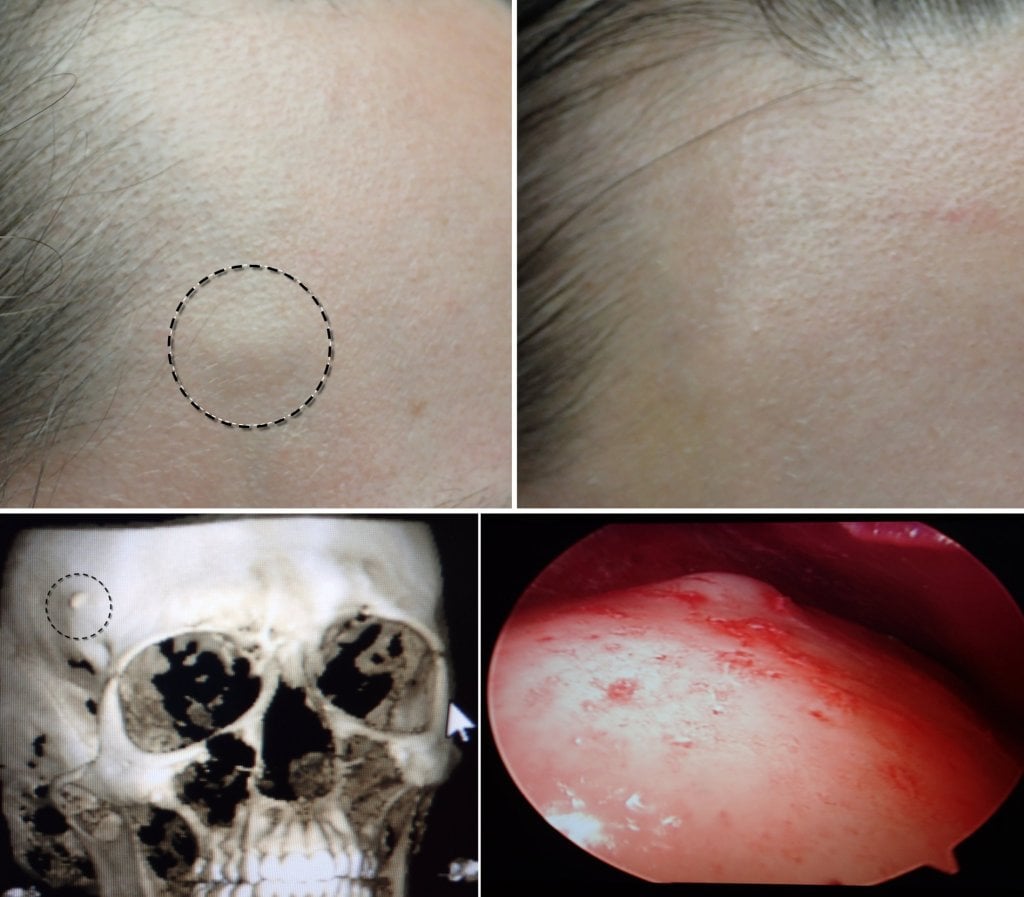
The CT scans below shows a typical forehead osteomas from the side and transverse views. The CT is performed to verify that the lesion is in fact a benign osteoma and not some other lesion that may communicate with deeper structures.


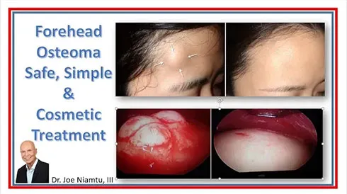
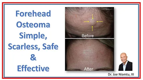
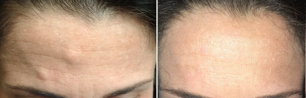
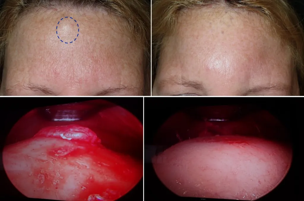
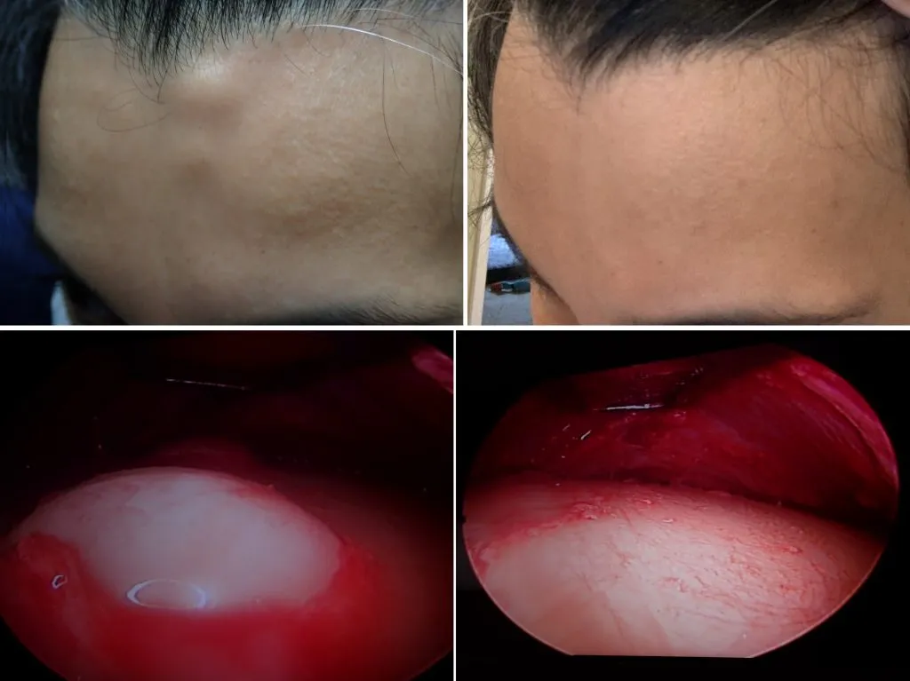
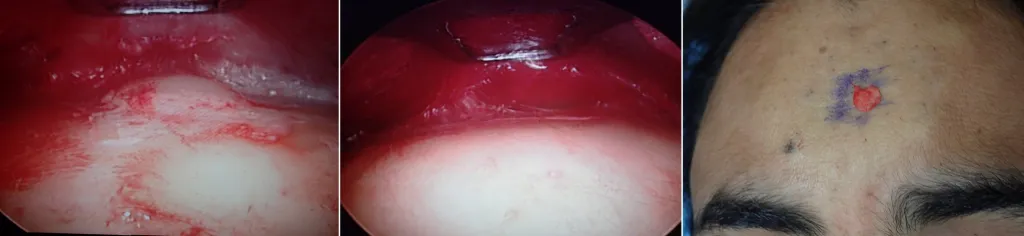
The images below show selected before and after cases of endoscopic osteoma removal.


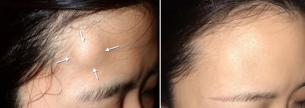

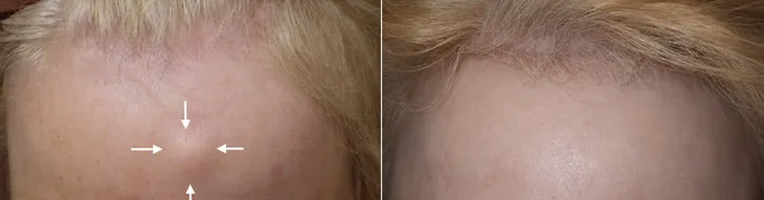


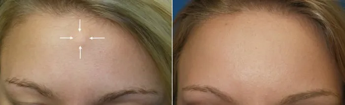


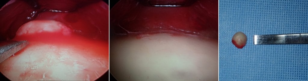


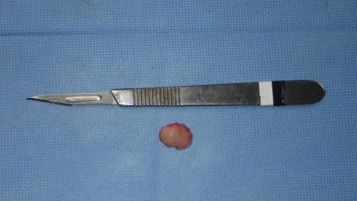
The above image shows the removed osteoma which is about the size of a small peach seed.
